Simple Compact Bone Diagram / Compact Bones vs. Spongy Bones - Difference and Comparison ... / This article explains the bone structure of the human body, using a labeled skeletal system diagram and a simple technique to memorize the names of all the the skeletal system is one of the important human body systems.
byAdmin-
0
Simple Compact Bone Diagram / Compact Bones vs. Spongy Bones - Difference and Comparison ... / This article explains the bone structure of the human body, using a labeled skeletal system diagram and a simple technique to memorize the names of all the the skeletal system is one of the important human body systems.. The compact bones form the hard exterior of the bones, whereas the spongy bones have several pores that are filled with nerves and blood vessels. Compact bone diagram compact bone diagram spongy bone diagram the histology guide cartilage bone ossification. The majority of growth during growth spurts is of the long bones. The foot bones shown in this diagram are the talus, navicular, cuneiform, cuboid, metatarsals and calcaneus. Over several more weeks or months, compact bone replaces spongy bone at the outer margins of the fracture and the bone is remodeled in response to strain (figure 6.5.2d).
This article explains the bone structure of the human body, using a labeled skeletal system diagram and a simple technique to memorize the names of all the the skeletal system is one of the important human body systems. Compact bone diagram simple diagram system. 5 bone tissue at brown mackie university. Compact bone is the denser, stronger of the two types of osseous tissue (figure 6.3.6). Start studying compact bone structure.
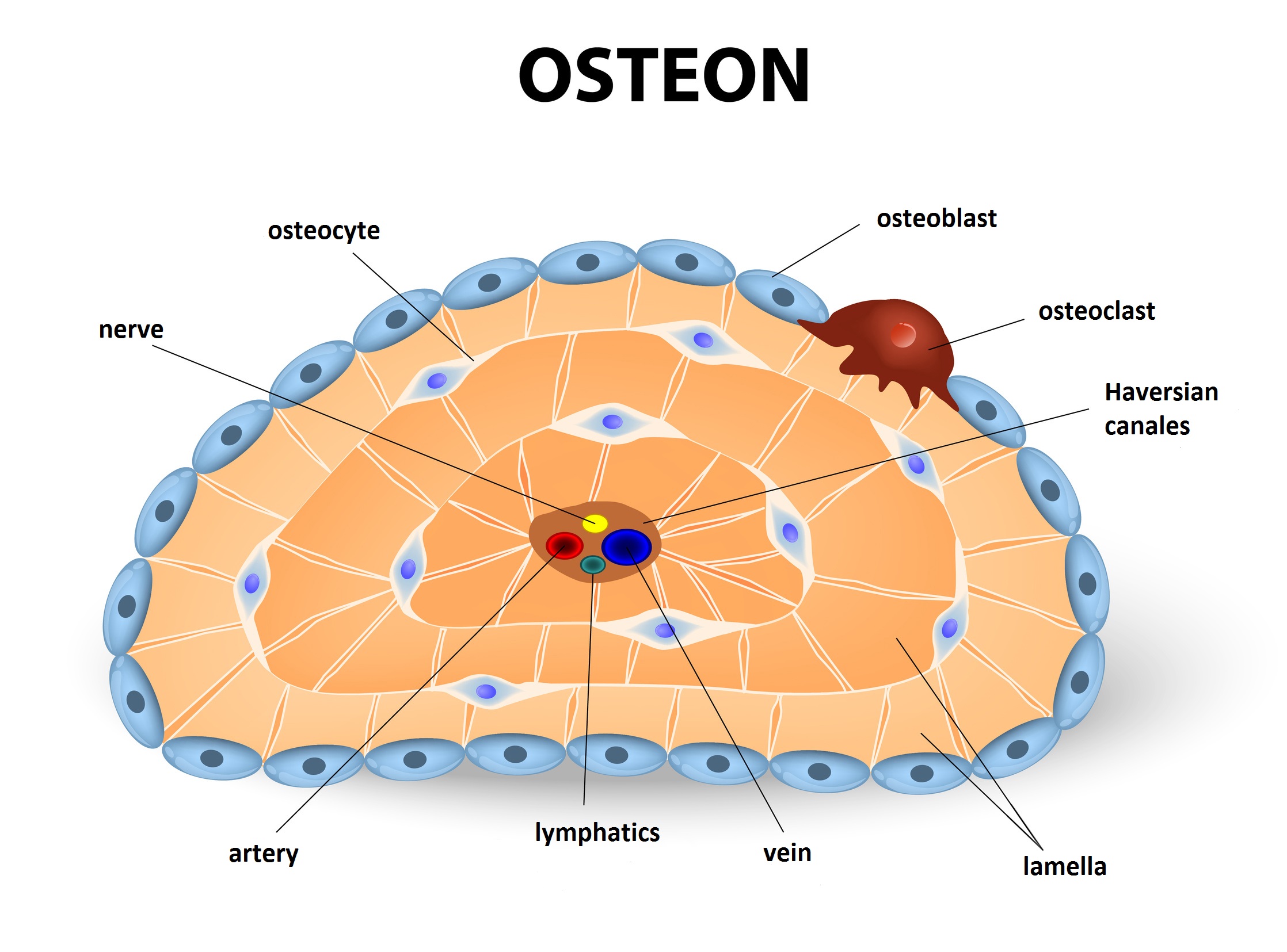
Bones: Fundamentals of anatomy for physicians | Lecturio from blog.lecturio.com However, they do contain osteons, which are like canals, providing passageways through the hard bone matrix. The microscopic structural unit of compact bone is called an osteon, or haversian system. Start studying leg bone anatomy. Bone is commonly classified according to its gross appearance as cancellous bone (bone with numerous, macroscopic interconnecting cavities, or trabeculae, also known as spongy or trabecular bone) or compact bone (dense lamellar bone without trabeculae), but both types have the same basic histological structure. In long bones, as you move from the outer cortical compact bone to the inner medullary cavity, the bone transitions to spongy bone. It makes up the outer cortex of all bones and is in immediate contact with the periosteum. Simple bone diagram wiring diagram. Animal cell structures functions diagrams simple animal cell drawing at getdrawingscom free for personal 50 diagram of the cell noibaiairporttransfer
However, they do contain osteons, which are like canals, providing passageways through the hard bone matrix.
It makes up the outer cortex of all bones and is in immediate contact with the periosteum. Compact bone, also called cortical bone, dense bone in which the bony matrix is solidly filled with organic ground substance and inorganic salts, leaving only tiny spaces (lacunae) that contain the osteocytes, or bone cells.compact bone makes up 80 percent of the human skeleton; Cartilage types, their location, bone types, classifications and god knows what else. It can be found under the periosteum and in the diaphyses of long bones, where it provides support and protection. Compact bone diagram simple diagram system. 5 bone tissue at brown mackie university. The compact bones form the hard exterior of the bones, whereas the spongy bones have several pores that are filled with nerves and blood vessels. Human skeleton long bones of arms and legs britannica. Related posts of compact bone diagram labeled anatomy of rib cage. The inset shows the lamellae of the. Compact bone definition compact bone, also known as cortical bone, is a denser material used to create much of the hard structure of the skeleton. Compact bone, as opposed to spongy bone, is made of cylindrical units, called osteons, that are tightly formed together. Cortical bone forms a dense cylinder down the shaft of the bone surrounding the central marrow cavity.
Bone is commonly classified according to its gross appearance as cancellous bone (bone with numerous, macroscopic interconnecting cavities, or trabeculae, also known as spongy or trabecular bone) or compact bone (dense lamellar bone without trabeculae), but both types have the same basic histological structure. Diagram of a typical long bone: Each osteon is composed of concentric rings of calcified matrix. The microscopic structural unit of compact bone is called an osteon, or haversian system. Shows compact (cortical) and cancellous (spongy) bone.
Compact Bone Diagram from www.dcicomp.com Illustration about compact bone, also called cortical bone, is the hard, stiff, smooth, thin, white bone tissue that surrounds all bones in the human body. It can be found under the periosteum and in the diaphyses of long bones, where it provides support and protection. Long bone diagram compact bone : Network wall socket wiring diagram. Ideal for studying bone structure and making function structure connections (as per ngss standards). (b) in this micrograph of the osteon, you can clearly see the concentric lamellae and central canals. The remainder of the bone is formed by cancellous or spongy bone. Each osteon is composed of concentric rings of calcified matrix.
Shows compact (cortical) and cancellous (spongy) bone.
Many tiny cells called osteocytes live in small spaces in the matrix and help to maintain the strength and integrity of the compact bone. Compact bone is made of a matrix of hard mineral salts reinforced with tough collagen fibers. Diagram of distinct morphological types of bone. Mcqs on cell, tissue, skeleton system and joint vipin vageria these pictures of this page are about:compact bone connective tissue diagram. However, they do contain osteons, which are like canals, providing passageways through the hard bone matrix. Illustration about compact bone, also called cortical bone, is the hard, stiff, smooth, thin, white bone tissue that surrounds all bones in the human body. The inset shows the lamellae of the. Learn vocabulary, terms, and more with flashcards, games, and other study tools. Bones are mostly made of the protein collagen, which forms a soft framework. Over several more weeks or months, compact bone replaces spongy bone at the outer margins of the fracture and the bone is remodeled in response to strain (figure 6.5.2d). Compact bone is the denser, stronger of the two types of osseous tissue (figure 6.3.6). Start studying leg bone anatomy. Microscopic anatomy bones medical massage physiology pediatric nursing medical knowledge medical anatomy anatomy tutorial structure of bone.
Pig bone diagram wiring diagram, femur bone diagram full human skeleton diagram femur simple compact bone diagram simple diagram system. Once healing and remodeling are complete a slight swelling may remain on the outer surface of the bone, but quite often, no external evidence of the fracture remains. Network wall socket wiring diagram. Diagramme schnell und einfach erstellen. However, they do contain osteons, which are like canals, providing passageways through the hard bone matrix.
structures of a long bone to label - Google Search ... from i.pinimg.com It can be found under the periosteum and in the diaphyses of long bones, where it provides support and protection. Long bones such as the femur contain two distinct morphological types of bone: Learn vocabulary, terms and more with flashcards, games and other study tools. About press copyright contact us creators advertise developers terms privacy policy & safety how youtube works test new features press copyright contact us creators. Osteons are the small units of which the hardest parts of human bones are made. The remainder is cancellous bone, which has a spongelike appearance with numerous large spaces and is found in the. It makes up the outer cortex of all bones and is in immediate contact with the periosteum. Some, mostly older, compact bone is remodelled to form these haversian systems (or osteons).
It seems confusing and misleading.
Bone is commonly classified according to its gross appearance as cancellous bone (bone with numerous, macroscopic interconnecting cavities, or trabeculae, also known as spongy or trabecular bone) or compact bone (dense lamellar bone without trabeculae), but both types have the same basic histological structure. Animal cell structures functions diagrams simple animal cell drawing at getdrawingscom free for personal 50 diagram of the cell noibaiairporttransfer Long bones such as the femur contain two distinct morphological types of bone: As seen in the image below, compact bone forms the cortex, or hard outer shell of most bones in the body. The remainder of the bone is formed by cancellous or spongy bone. Pig bone diagram wiring diagram, femur bone diagram full human skeleton diagram femur simple anatomy, colored ear diagram for kids bone labeled of the eye to label compact bone diagram simple diagram system. Illustration about compact bone, also called cortical bone, is the hard, stiff, smooth, thin, white bone tissue that surrounds all bones in the human body. Microscopic anatomy bones medical massage physiology pediatric nursing medical knowledge medical anatomy anatomy tutorial structure of bone. It makes up the outer cortex of all bones and is in immediate contact with the periosteum. This article explains the bone structure of the human body, using a labeled skeletal system diagram and a simple technique to memorize the names of all the the skeletal system is one of the important human body systems. Pig bone diagram wiring diagram, femur bone diagram full human skeleton diagram femur simple compact bone diagram simple diagram system. Compact bone is the denser, stronger of the two types of osseous tissue (figure 6.3.6). Under the periosteum is a thin layer of compact bone (often called cortical bone).
Once healing and remodeling are complete a slight swelling may remain on the outer surface of the bone, but quite often, no external evidence of the fracture remains compact bone diagram. Compact bone diagram compact bone diagram spongy bone diagram the histology guide cartilage bone ossification.